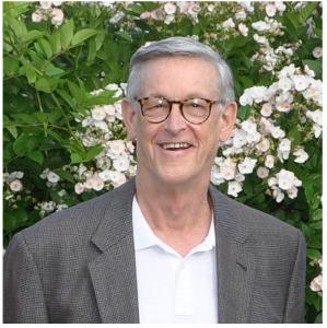
David Rowe
Professor
Department of Reconstructive Sciences
David Rowe received the M.D. degree from the University of Vermont, Burlington, VT, in 1969. He is currently a Professor in the Department of Reconstructive Sciences, University of Connecticut Health, Farmington, CT, where he has been Physician Health Services Chair in human genetics since 1992. He is also the Director of Regenerative Medicine and Skeletal Development, School of Dental Medicine, Farmington.
Dr. Rowe was initiated into skeletal biology as a Clinical Associate in the Dental Institute of NIH in the biochemistry laboratory of Drs. Karl Piez and George Martin. At the time the primary structure of type I collagen was being determined and steps of processing procollagen to collagen were being discovered. It was during that time that the first gene mutations affecting collagen that lead to certain forms of Ehlers Danlos and Osteogenesis Imperfecta were identified. Thus Dr. Rowe has observed and participated in the progression of genetics, molecular biology, stem cell and regenerative medicine develop as it applies to human skeletal health. His laboratory effort has been primarily based on the mouse as an effective surrogate for the human diseases of the skeleton.
As the genetically manipulated mouse became the standard for studying human disease, Dr. Rowe began to focus on how to develop sensitive, low cost and high throughput technologies for characterizing the histological features of skeletal tissues. The sensitivity and specificity of fluorescent based imaging because increasingly evident and methods had to be developed to take advantage of these properties. Working closely with Dr. Xi Jiang, a procedure for capturing intact sections of frozen non-decalcified skeletal tissue because the base procedure for histological staining and imaging that is used through out our mouse based studies. The ability to co-localize the structure of the mineralized tissue, its associated mineralization lines, plus the presence of osteoblasts and osteoclasts all on the same section brought efficiency and histological registration to a new level of resoluton.
At the same time that the histological advances were being made, Dr. Rowe and Shin had been collaborating in developing computational tools for organizing and interpreting microarray studies. When Dr. Hong, who was trained in image analysis, joined Dr. Shin’s laboratory, an opportunity was realized that the fluorescent signals from the new histology could be recognized by the computer. Over the ensuing 2-3 years, the algorithms that discriminate, calculate and display the relationship of the signals to the bone surface were developed. As this computer-based analysis became automated, we realized that his program could be expanded to a wide variety of skeletal tissues and biological processes that involve mineralized tissue formation and turnover.
The opportunity to demonstrate that our histological acquisition and image analysis could be expanded to a larger dataset became a reality with the funding of the KOMP2 program for skeletal phenotyping. Dr. Cheryl Ackert Bicknell joined our group effort and provided the genetic expertise and access to the mice that were being evaluated in the KOMP pipeline at the Jackson Laboratory. This source of genetically characterized mice on a highly defined genetic background give us the opportunity to demonstrate that skeletal phenotyping can be performed in a objective and consistent manner, and that the information can be presented to the public in a organized, searchable and biologically relevant manner. Thus, the goal of Dr. Rowe’s effort in KOMP is that the concept can be expanded to other sources of genetically modified mice and to other mineralizing skeletal tissues (mineralizing cartilage, dentition, and calvarial sutures).

| rowe@uchc.edu | |
| Phone | 860-679-2461 |
| Mailing Address | Department of Reconstructive Sciences, 263 Farmington Avenue, MC 3705, Farmington, CT 06030 |
| Office Location | L7009 |
| Campus | UConn-Farmington |